Research
Goal of our Research
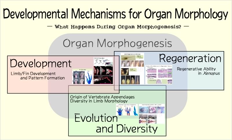
Our research interest is in understanding the molecular and cellular mechanisms underlying organ morphogenesis in vertebrates. Each organ has a characteristic shape that is required for its proper function. During development, the structure of an organ is produced by intrinsic and genetic programs in the embryo. Our primary goal is to elucidate the developmental programs and the subsequent behavior of cells and tissues that generate distinct morphology of organs. Knowledge obtained through these studies will be useful for considering our further interest, that is, issues of the morphological diversity of organs, which is caused by modifications and alterations of the developmental programs during evolution.
We are also investigating the mechanisms of organ regeneration. Although appropriate morphogenesis is essential to regenerate functional replica of lost organs, little attention has been paid to this in recent regeneration studies such as studies on stem cell biology. Result of our studies on organ morphogenesis during development and regeneration will contribute to the establishment of a scientific basis for regenerative medicine.

(Click image to enlarge.)
Limb Development
During vertebrate development, limbs arise as swellings called limb buds from the lateral plate mesoderm in the ventrolateral body wall. The limb-forming regions are thought to be determined by the surrounding tissues and their positions are precisely defined. Once the limb bud has formed, morphogenesis occurs along three axes, antero-posterior, dorso-ventral and proximo-distal axes. Some organizing centers for morphogenesis along each axis are known to exist in the limb bud (such as the ZPA, AER, and non-AER ectoderm). We focus on two main questions for this field of research. One is how limbs are formed at specific positions of the body wall. The other is how limb bud cells translate positional information along the axes into limb morphology. Results of these studies will provide insights into the general mechanisms of organ morphogenesis during embryonic development.

(Click image to enlarge.)
Limb Regeneration
Urodele amphibians such as the newt and axolotl can completely regenerate their limbs throughout their life. In contrast, the ability of the anuran amphibian Xenopus laevis to regenerate limbs drastically changes as it gets older. Although young tadpoles can regenerate complete limbs after amputations of developing limb buds, older tadpoles have almost lost the ability for regeneration. On the other hand, froglets (young adults shortly after metamorphosis) can regenerate only a hypomorphic cartilage spike. The spike has no joint and bifurcation, suggesting that it fails to regenerate the skeletal pattern along the axes of the limb. Understanding the causes of reduction of their regenerative ability will clarify the requisites for complete regeneration of organ morphology and functions. In the future, we will explore the reasons why mammals (and other amniotes) have severely limited ability to regenerate organs.

(Click image to enlarge.)
Evolution and Diversity of Vertebrate Appendages
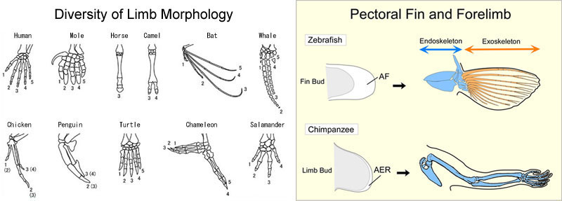
Tetrapod limbs evolved from paired (pectoral and pelvic) fins of ancestral fishes and altered their morphology to adapt to their respective environments. For example, forelimbs of birds became wings for flying and marine mammals such as dolphins and seals changed their limbs to paddle-like flippers to swim like fish. While developmental processes of these various limbs are thought to share common basic mechanisms, there are certain differences in the mechanisms that may cause their divergent morphology. What are the differences? Are there some rules or constraints in the alteration of the mechanisms? We will attempt to answer these questions.
Another major issue in this field is fin-limb transition. Although fish fins and tetrapod limbs share many common mechanisms of development, their morphologies in adult animals are quite different. Paired fins have extended exoskeletons (fin rays) in the distal part and poorly developed endoskeletons (radials) in the proximal part. In contrast, the distal part of limbs has well developed endoskeletons of the autopod (wrist/ankle and hand/foot) instead of the exoskeletal fin rays. We are investigating the mechanisms that account for these morphological differences from the viewpoint of developmental biology.

(Click image to enlarge.)
